Quantitative Microscopy
The stereological methods such as Cavalieri method and method based on cycloidal arcs were used to estimate volume fractions of the spongiosa and the compacta and surface density of myocytes in the spongiosa. The parameters were estimated for heart arrested in diastole as well as in systole.
Materials and Methods
From two carps (Cyprinus carpio L., weight 1.7 kg), beating hearts were
explanted and connected to a Langendorff set up (Fig. The Langendorff set up with treatment solutions.),
[sipkema-et-al-1998]). Heart #1 was treated with a solution containing
 (2-3 mmol/l) and thus arrested in systole. Heart #2
was treated with a
(2-3 mmol/l) and thus arrested in systole. Heart #2
was treated with a  solution (20 mmol/l) and arrested in
diastole, see Figs. The Langendorff set up with treatment solutions. and The hearts of the carps..
solution (20 mmol/l) and arrested in
diastole, see Figs. The Langendorff set up with treatment solutions. and The hearts of the carps..
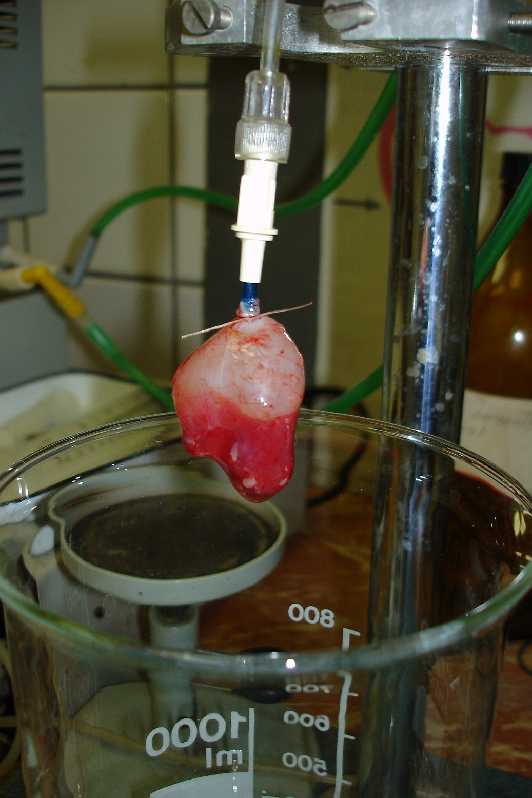
The Langendorff set up with treatment solutions.
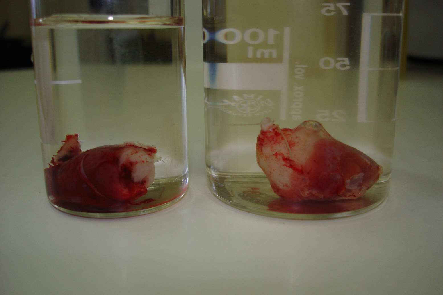
The hearts of the carps.
Both hearts were then perfused with 4% buffered formalin solution, embedded in
paraffine and cut using the microtome Leica RM 2135 (Leica Microsystems GmbH,
Germany) to serial sections with thickness 6  . Each
 section (86 sections in heart #1, 98 sections in heart #2)
was saved on a slide, stained with Verheoff’s hematoxylin and green trichrome
and used for stereological quantification. Only the ventricle was
investigated. The cutting plane was oriented perpendicularly to the long axis
of the ventricle. The whole sections were scanned on a flatbed scanner with
1200 dpi resolution. Every second section was then also photographed with a
4
section (86 sections in heart #1, 98 sections in heart #2)
was saved on a slide, stained with Verheoff’s hematoxylin and green trichrome
and used for stereological quantification. Only the ventricle was
investigated. The cutting plane was oriented perpendicularly to the long axis
of the ventricle. The whole sections were scanned on a flatbed scanner with
1200 dpi resolution. Every second section was then also photographed with a
4  objective.
Quantitative Microscopy
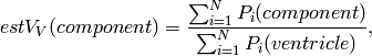
We assessed the volume fraction of compacta  and spongiosa
  within the
ventricle using the point-grid test system placed on the micrographs of the
tissue according to (see [howard-reed-1998]):
within the
ventricle using the point-grid test system placed on the micrographs of the
tissue according to (see [howard-reed-1998]):
where  was either compacta or spongiosa, and
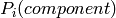 was the number of points of the test system lying on
transections on
was the number of points of the test system lying on
transections on  -th section and was the number
of points within reference space, namely points that intersected the heart on
-th section and was the number
of points within reference space, namely points that intersected the heart on
 section, see Figs. The point-grid test system (compacta).,
The point-grid test system (spongiosa)..
section, see Figs. The point-grid test system (compacta).,
The point-grid test system (spongiosa)..
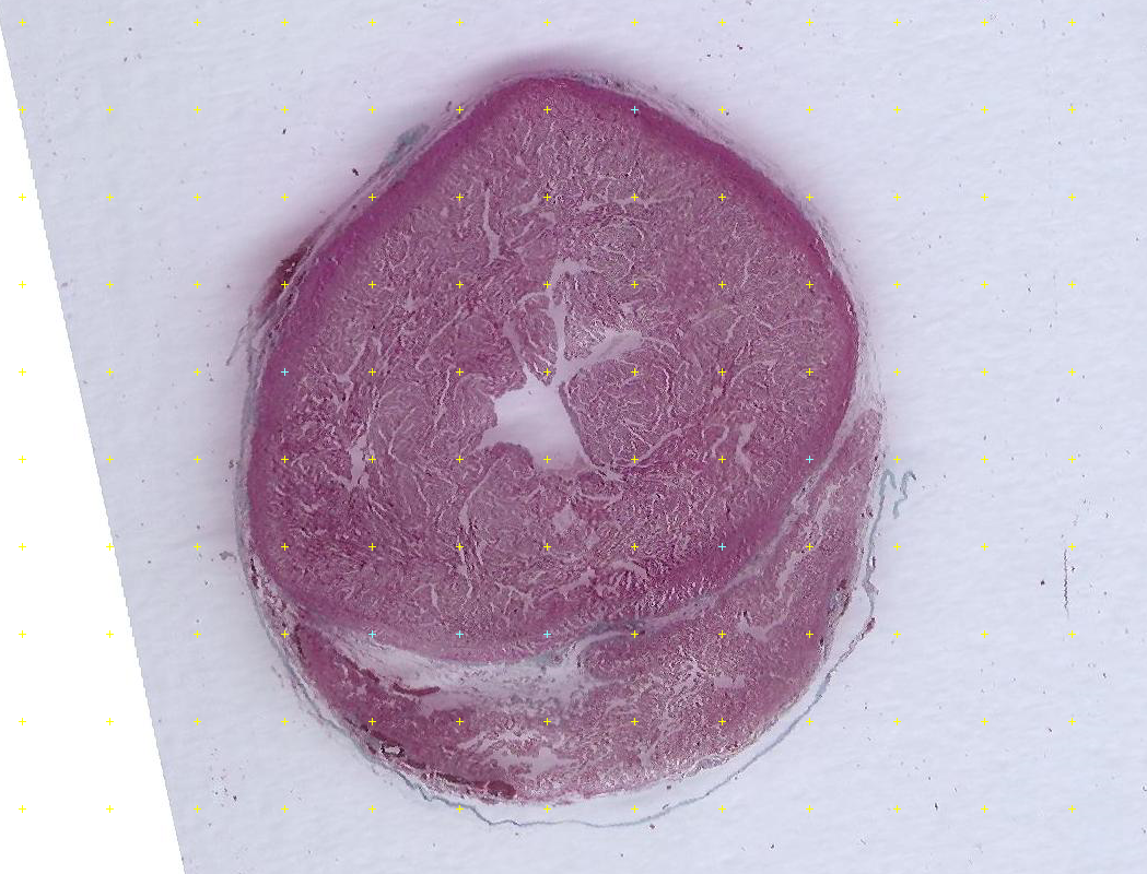
The point-grid test system (compacta).
The point-grid test system (spongiosa).
For estimating the volume fraction of the spongiosa within the wall of the
ventricle , the microscopic
spaces among the trabeculas of the spongy myocardium were considered as part of
the spongiosa, because they were undistinguishable at the level of whole
scanned sections used for this estimation.
The spongiosa appeared as a complex of branching and anastomosing trabeculae
with a considerable surface. Hypothetically, the geometry of the surface might
have differ in systole and diastole. Therefore, surface density of spongiosa
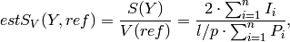
 was assessed as follows (see [howard-reed-1998]):
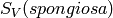
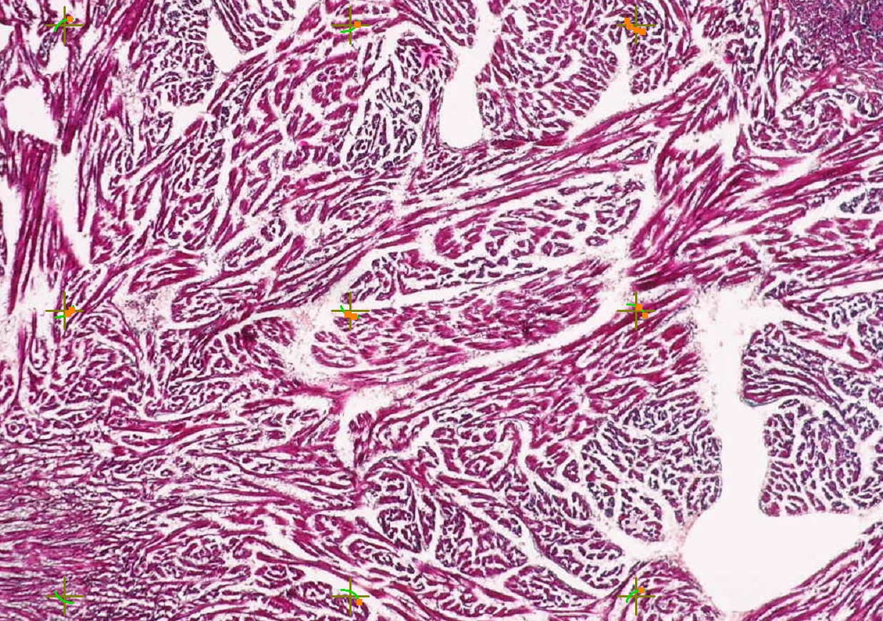
where 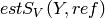 was the surface density of object
  (spongiosa) within the reference volume
  (spongiosa without
cavities),
(spongiosa without
cavities),  was surface area of object ,
  was
the known length of cycloidal arc per point,
was
the known length of cycloidal arc per point,  was the number of
intersections between circular arcs and the boundary of object and
was the number of
intersections between circular arcs and the boundary of object and  was the number of points hitting the reference space, see
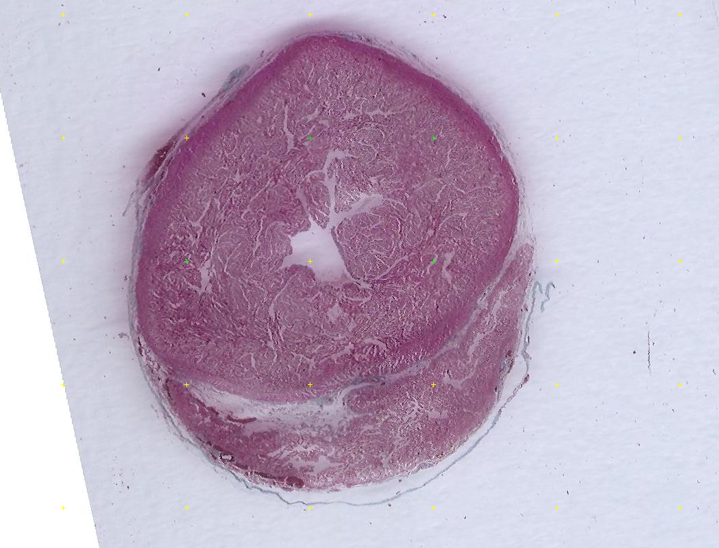
Figs. The system of cycloidal arcs., The point-grid test system (reference volume)..
was the number of points hitting the reference space, see
Figs. The system of cycloidal arcs., The point-grid test system (reference volume)..
The system of cycloidal arcs.
The point-grid test system (reference volume).
Results
The results are shown in the following table:
| parameter | heart #1 (systole) | heart #2 (diastole) |
|---|---|---|
| 1. |
20.70% | 19.60% |
2.  |
58.93% | 62.79% |
3.  |
0.35 | 0.31 |
4.  |
0.0949  |
0.1466 |
5.  |
37.80% | 28.56% |
6.  |
41.51% | 51.84 |
7.  |
0.55 | 0.69 |
The volume fraction of the compacta within the ventricle (parameter 1) was independent of the phase of the heart cycle, as the compact layer of the myocardium was apparently incompressible. The volume fraction of the spongiosa in the ventricle (parameter 2) is not to be interpreted as this estimation did not take into account the microscopic spaces among the trabeculae of the myocardium. Therefore, also the ratio between compacta and spongiosa (parameter 3) offered no suitable interpretation, when the microscopic spaces among the spongy trabeculae were not distinguished from the trabeculae and thus falsely increasing the volume of the spongiosa.
However, the surface density of spongiosa (parameter 4) was considerably lower in systole than in diastole. In other words, the systolic myocardium had more collapsed spongy trabeculae than the diastolic myocardium. This parameter demonstrated that the same volume of the spongiosa had a lower surface in systole than in diastole. This corresponded to the higher volume fraction of the spongiosa within the systolic ventricle (parameter 5) and to the lower volume fraction of heart cavities within the systolic ventricle (parameter 6). As demonstrated with the parameter 7, the ratio between the volume of compacta and the volume of spongiosa is higher in systole, which corresponds to the increased volume fraction of spongiosa in the collapsed ventricle during the systole (parameter 5).
| [howard-reed-1998] | (1, 2) Howard, C.V., Reed, M.G., 1998. Unbiased Stereology: Three Dimensional Measurement in Microscopy. 1st edn. Royal Microscopical Society and Springer-Verlag, New York, 246 p. |
| [sipkema-et-al-1998] | Sipkema P., Takkenberg J.J.M., Zeeuwe P.E.M., Westerhof N. (1998): Left coronary pressure–flow relations of the beating and arrested rabbit heart at different ventricular volumes. Cardiovascular Research 40: 88–95. |